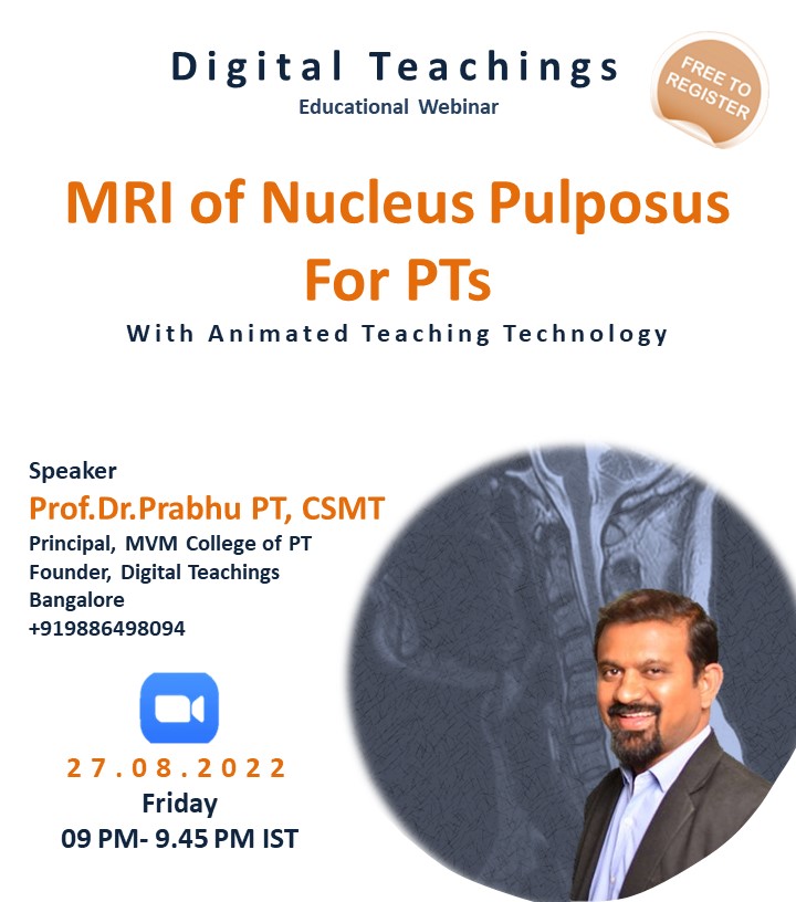
Welcome to FRCPT Continuing Educational Webinar.
MRI protocol for identifying for nucleus pulposus and annular fibrosus is quite simple. T1 & T2-weighted images are routinely performed in the evaluation of the spine12.

Basically we rely on the sagittal T1W & T2W images and correlate the findings with the transverse T2W images6.
It is easy to analyze the disc by comparing T1W & T2W MRI Images. The sagittal T1W images give you the basic picture of Intervertebral disc.
Workshop Contents
Comparison of T1 & T2 Weighted MRI
Nucleus & Annulus Difference in T1w & T2w
Cortical Bone in MRI
Disc & Bone Cortex in T1W MRI
Bone Cortex & Annulus Fibrosus in T2w MRI
Normal Disc in Sagittal T1 & T2 Weighted MRI
Age related Radiological Changes of Nucleus Pulposus
Nucleus Grading System
Disc in T2w Axial View

Know more about FRCPT Course.


Add Comment