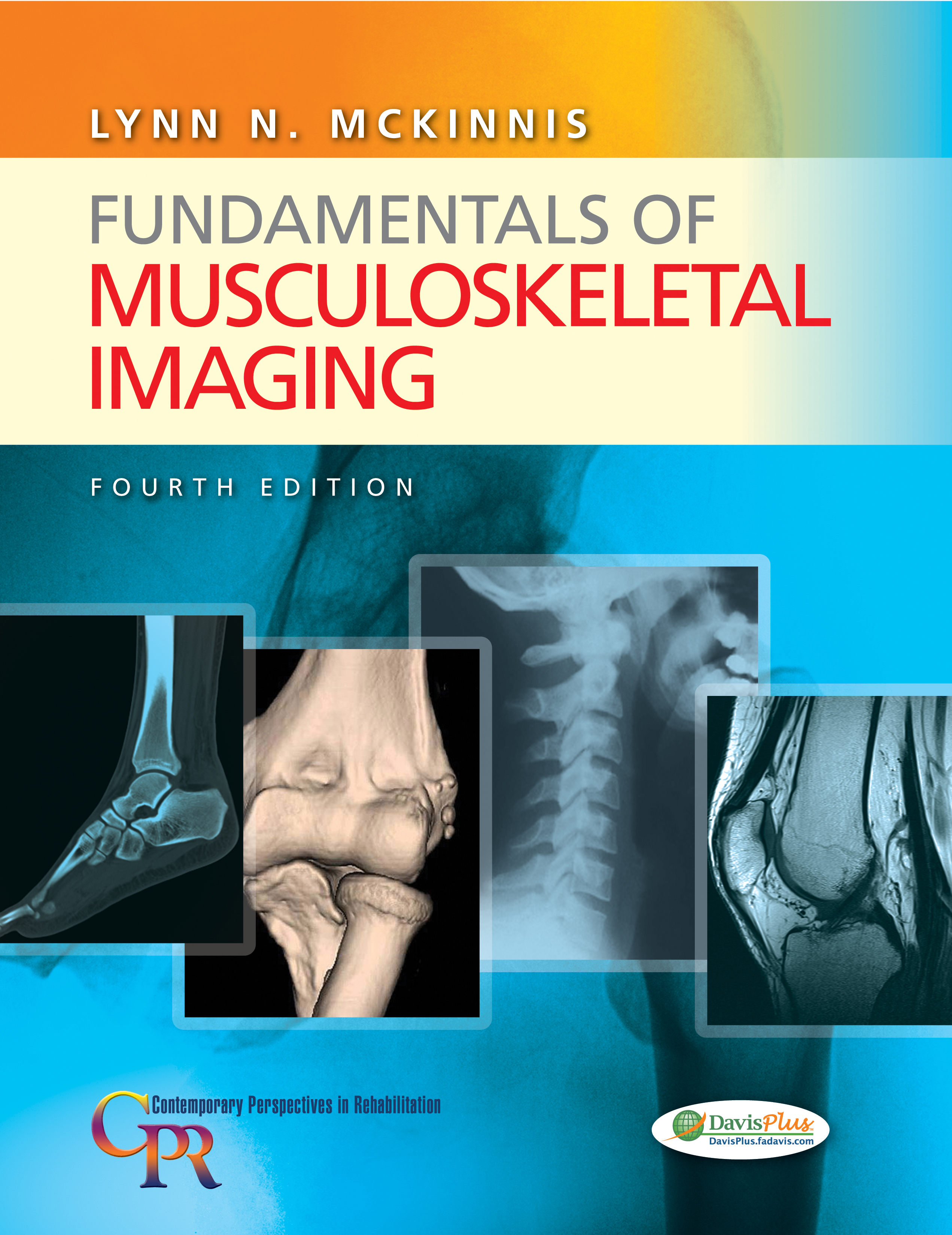
Excerpts from
“Fundamentals of Musculoskeletal Imaging”

Lynn McKinnis
By Lynn McKinnis PT, DPT
Recipient of
APTA’s Helen J. Hislop Award for Outstanding Contributions to Professional Literature – 2009
Traditionally the field of imaging has been the domain of the physician. Physiotherapists have generally excluded themselves from interaction with imaging. This self-built professional boundary evolved from a misconception that information gained from viewing images was pertinent only to the medical diagnosis and therefore pertinent only to the physician. Studying the patient’s diagnostic images was rarely considered, and simply reading the radiologist’s “x-ray report” was accepted as sufficient. It was a long-held erroneous belief of Physiotherapists that, even if images contained a wealth of information to enhance patient treatment, Physiotherapists were incapable of finding it on their own (1).
Traditions change.
Later, Physiotherapists have discovered that their knowledge of functional anatomy is an excellent foundation for visually comprehending diagnostic images as well as correlating clinical findings with imaging findings. The inclusion of diagnostic imaging courses in educational and professional settings is giving physiotherapists the confidence to dialogue with radiologists, gain relevant information from the radiologist’s report, and most significantly, view diagnostic images with their own eyes.
Traditions change slowly.
Although it is accepted as logical that Physiotherapists need to be aware of the patient’s medical diagnosis, it is considered novel by some that these Physiotherapists view diagnostic images themselves. For others the idea is more than novel—it can appear threatening if it is mistakenly perceived by physicians as a move toward diagnosis or second-guessing the diagnosis or if it is mistakenly perceived by clinicians as either an opportunity or a responsibility to do just that. Professional collaboration to enhance the quality of patient care is the single most important goal.
Traditions change for good reasons.
Why do Physiotherapists need to view diagnostic images? (1).
1. A more comprehensive evaluation is obtained. The success of rehabilitation depends on the effectiveness of the Physiotherapist’s evaluation. The more thorough the evaluation, the more substance the Physiotherapist has on which to build the rehabilitation program. Many of the Physiotherapist’s evaluation tools—observation, palpation, goniometry, manual muscle testing, ligamentous stress testing, joint-end feels, joint mobility testing— are dependent on the Physiotherapist’s own perceptive skills and have an inherent degree of subjectivity and limitation.
Imaging can provide an objective, visual aspect to the evaluation that makes the expertise of the physiotherapist more comprehensive. Supplementing the initial evaluation and re-evaluations with musculoskeletal images increases the Physiotherapist’s awareness of the patient in an added dimension. The Physiotherapist’s knowledge of functional anatomy becomes more dynamically effective by allowing direct visualization of the processes of growth, development, healing, disease, and dysfunction.
2. The information the Physiotherapist seeks is often of a different nature than the information the physician seeks and of a different nature than may be described in the radiologist’s report. For example, a physician needs to know whether a fracture of the distal radius that has united with a malunion deformity is clinically stable; if so, the cast can be removed and the patient can be sent for rehabilitation. The Physiotherapist, however, also needs to know the severity and configuration of the malunion deformity. By viewing the patient’s radiographs, the Physiotherapist becomes aware of how the adjacent joints of the hand, wrist, forearm, and elbow have the potential to be affected by the deformity. The Physiotherapist’s treatment goals may thus be modified from obtaining full premorbid range of motion to obtaining a lesser degree of motion, adequate for function but minimizing the abnormal joint arthrokinematics that may accelerate degenerative changes in the joints.
Guidelines:
The growth of direct access, with physical therapists assuming primary care roles, and the transition to the doctor of physical therapy degree are among the changes related to practice and education in the physical therapy profession that have increased physical therapists’ need to be knowledgeable about diagnostic imaging.
The APTA documents “Vision Statement 2020,” “Guide to Physical Therapist Practice,” “A Normative Model of Physical Therapist Professional Education,” and “Orthopaedic Physical Therapy Description of Specialty Practice” all contain language supportive of physical therapists’ use of diagnostic imaging information in clinical practice” (2,3,4,5).
Access to imaging is crucial for medical screening in primary care practice, because diagnostic imaging sometimes provides diagnostic information not available through the interview and physical examination. For example, in the case of a typical ankle inversion injury, a radiograph may be diagnostic for an avulsion fracture, which necessitates referral to a physician for management, or it may rule out a fracture, confirming that the soft tissue injury can be managed by the physical therapist. Furthermore, in the case of a more serious ankle sprain with instability, the physical therapist may conclude that the radiograph is not useful in evaluating ligaments and may suggest that MRI be performed to define which tissues are involved. As part of any interdisciplinary health-care team, physiotherapists must be able to confer with physicians about diagnostic imaging, suggest appropriate diagnostic imaging to the patient’s primary care physician, and refer directly to the radiologist if needed (1).
Direct referral to a radiologist for diagnostic imaging may be efficient and cost saving method. Physiotherapists are expected to refer patients to other physician specialists when necessary (such as orthopedic surgeons, internal medicine specialists, and neurologists); radiologists would not seem to be an exception (1).
Legal Aspects around the World:
In the United States, physical therapists practicing in the military, in private practice, in health maintenance organizations, in the Veterans Administration, and in other settings may all assume primary care roles, thus necessitating knowledge of and access to diagnostic imaging. (6, 7). The physiotherapists were able to reduce the number of radiological examinations by more than 50% in a population of 2,117 patients with low back pain (8), The Physiotherapists’ clinical diagnostic accuracy, as compared with MRI findings, is similar to that of orthopedic surgeons and significantly greater than that of non-orthopedic providers (9).
Physiotherapists in Australia, New Zealand, and the United Kingdom all have the ability to directly obtain diagnostic imaging for patients within their roles as primary care providers (10-13). Almost 74% of physiotherapists do refer patients for radiographs at an overall annual rate of 8.3 referrals per physiotherapist (11).
Physiotherapists in New Zealand were recognized by the Accident Compensation Commission (ACC) in 1999 as providers able to refer patients for both conventional radiographs and ultrasound imaging (13).
In United Kingdom, Physiotherapists who are registered as extended scope practitioners (ESPs) possess the ability to refer patients for diagnostic imaging (12).
The radiologist bears the ultimate responsibility for the radiological diagnosis. In brief, the orthopedic radiologist attempts to diagnose an unknown disorder, demonstrate the exact location, identify the distribution of the lesion in the skeleton, gain pertinent information for the surgeon, and monitor the response of the lesion to medical intervention. Once a disorder is identified in this way, the function of diagnostic imaging is complete as far as the physician is concerned. A physiotherapist, however, may use imaging studies for purposes other than diagnosis but related to rehabilitation of the diagnosed pathology. Because the radiology report is written to serve the physician’s decision-making needs, information specific to the physical therapist’s intervention planning is not usually provided. The ability to comprehend the image itself, then, is valuable for the physical therapist.
Physiotherapists are typically responsible for restoring normal movement to a joint that has been directly or indirectly affected by a disorder. Before choosing specific treatment techniques to address the movement limitations, physical therapists want to know:
1. What barriers to normal movement might exist at the joint? Surgical fixation devices, loose bodies, and excessive callus can all be problematic.
2. Which interventions (passive, active, resistive maneuvers) for restoring movement are safe and appropriate at any given point in a patient’s rehabilitation?
3. How much stress applied to healing tissues will promote the return of normal function without compromising optimal healing?
4. Where should the limb be stabilized to avoid movement at a fracture site? Can an adjacent joint be mobilized without endangering the fracture site?
5. How much weight-bearing is safe at this time?
Sometimes viewing available imaging studies can help answer these questions. If not, consultation with the physician regarding the images may answer them. Even a consultation, however, requires that the physical therapist understand an image well enough to discuss it. So universities should include basic radiology program in the Physiotherapy course. The preferred training method in U.S military is a 2-week basic radiology course as part of Physiotherapy university program (6); same could be followed in other universities to have better physiotherapy practice.
These are the starting points for the relatively new idea of a frequent intersection between the fields of Physiotherapy and Imaging. The future holds potential for other synergies that can advance the scopes of both fields. The goal of this workshop is to provide an understanding of Spinal imaging fundamentals so that the content, possibilities, and limitations of diagnostic images can be appreciated.
References:
1. McKinnis, Lynn N. (1959). Fundementals of Musculoskeletal imaging. Third Edition. F A Davis, Philadelphia.
2.APTA: 2007–2008 Fact Sheet, Physical Therapist Education Programs. APTA, Alexandria,VA,May 2008.
3. Rothstein, JM (ed): Guide to Physical Therapist Practice, ed. 2. Phys Ther 81:9, 2001.
4. APTA: A Normative Model of Physical Therapist Professional Education: Version 2004. APTA, Alexandria,VA, 2004.
5. American Board of Physical Therapy Specialties Specialty Council on Orthopaedic Physical Therapy: Orthopaedic Physical Therapy Description of Specialty Practice. APTA, Alexandria,VA, 2002.
6. Dininny, P: More than a uniform: The military model of physical therapy. PT Magazine 3(3):40, 1995.
7. Greathouse, DG, et al: The United States Army physical therapy experience: Evaluation and treatment of patients with neuromusculoskeletal disorders. J Orthop Sports Phys Ther 19:261, 1994.
8. James, JJ, and Stuart, RB: Expanded role for the physical therapist: Screening musculoskeletal disorders. Phys Ther 55:121, 1975.
9. Moore, JH, et al: Clinical diagnostic accuracy and magnetic resonance imaging of patients referred by physical therapists, orthopaedic surgeons, and nonorthopaedic providers. J Orthop Sports Phys Ther 35:67, 2005.
10. Littlejohn, F, Nahna, M, Newland, C, et al:What are the protocols for imaging referral by physiotherapists? N Z J Physiother 34(2):81, 2006.
11. Australian Physiotherapy Association: Physiotherapists Diagnostic Imaging Referral Patterns, Presented to the Department of Health and Aging. Australian Physiotherapy Association, August 2004.
12. The Chartered Society of Physiotherapy: Chartered physiotherapists working as extended scope practitioners. Information paper No. PA29. London, 2002.
13. Scrymgeour, J:Moving on: A history of the New Zealand Society of Physiotherapists, Inc. 1973–1999.Wellington, The New Zealand Society of Physiotherapists Inc., 2000.

